


What Is This Bump? A Guide to Common Skin Lesions and When to Be Concerned
- Dec 18, 2025
- 5 min read
Finding a new bump, spot, or patch on your skin can be unsettling. Most of the time, these changes are harmless — but occasionally they can signal something more serious. At MedHub, we believe in informed awareness and prompt assessment by a trained dermatologist using specialised tools like a dermatoscope. Ultimately, histology (a biopsy examined under a microscope) remains the gold standard for diagnosis.
Understanding Skin Lesions
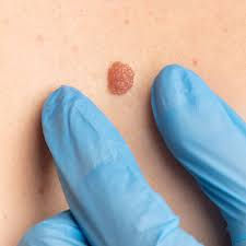
A skin lesion is any area of the skin that looks different from the surrounding tissue. Lesions can be flat or raised, pigmented or skin-coloured, smooth or crusty, and may appear anywhere on the body.
Here’s a quick guide to common types — and clues that suggest when you should get them checked:
Common Benign (Non-Cancerous) Skin Lesions
🟤 1. Seborrhoeic Keratosis
These are very common, especially in middle-aged and older adults.
Appearance:
“Stuck-on” waxy or wart-like bump
Creamy, tan, brown or black
Rough or smooth surface
When it’s usually harmless:No change in size or colour over time, no bleeding or itching.
⚪ 2. Milia
Tiny white or yellow cysts often seen on the face.
Appearance:
Small, firm, pearly bumps
Often around eyes or cheeks
Usually harmless:Simple reassurance or cosmetic removal is all that’s needed.
🟠 3. Dermatofibroma
Firm, small nodules most common on legs.
Appearance:
Dome-shaped, firm when pinched
Often darker than surrounding skin
Typical behaviour:Stable in size and texture; benign.
💡 4. Lipoma
Soft fatty lumps under the skin.
Appearance:
Soft, mobile under the finger
Usually painless
Benign:Can enlarge slowly over years; removed if bothersome.
Things That Might Be Concerning
⚠️ 5. Atypical (Dysplastic) Mole
These are benign but have irregular features.
Appearance:
Asymmetric shape
Uneven colour
Border irregularity
Larger than a pencil eraser
Concern:These may have a higher risk of transforming into melanoma.
🔴 6. Basal Cell Carcinoma (BCC)
A common skin cancer that doesn’t usually spread but can damage surrounding tissue.
Red flags:
Pearly, translucent bump
Small blood vessels visible on surface
Tendency to bleed or ulcerate
⚫ 7. Squamous Cell Carcinoma (SCC)
Can be more aggressive than BCC.
Red flags:
Rough, scaly patch
Ulcer or persistent sore
Tender or growing
🖤 8. Melanoma
The most dangerous form of skin cancer if not caught early.
Remember the ABCDEs:
Asymmetry
Border irregular
Colour variation
Diameter increasing
Evolving over time
Any mole changing rapidly — growing, itching, bleeding, darker in parts — needs urgent evaluation.

The Role of Dermatoscopy
A dermatoscope is a handheld microscope that lets a dermatologist examine structures beneath the surface of the skin. It helps differentiate benign lesions from suspicious ones far better than the naked eye alone.
Why this matters:
Some melanomas can look like harmless freckles.
Atypical moles may have subtle features only visible dermatoscopically.
Early skin cancers may be diagnosed before they spread.
Dermatoscopy improves diagnostic accuracy and helps decide which lesions need removal or biopsy.
Histology: The Definitive Diagnosis
While clinical evaluation and dermatoscopy guide decision-making, histology — examining a tissue sample under a microscope — provides a definitive diagnosis.
A biopsy may be recommended when:
A lesion looks unusual or is changing
Dermatoscopy shows concerning patterns
There is persistent bleeding, ulceration, or rapid growth
Histology tells us exactly what the cells are — be they benign, precancerous, or malignant.
When to See a Dermatologist
Seek specialist assessment if you notice:
✔ A new lesion that appears suddenly
✔ A bump that is rapidly growing
✔ A mole changing shape, colour, or size
✔ Lesions that bleed, itch, crust, or don’t heal
✔ Multiple atypical moles or a personal history of skin cancer
✔ Anything that simply “feels wrong”
Your GP can refer you to dermatology, but if your concern is urgent — for example, melanoma suspicion — ensure you communicate that clearly.
Take‐Home Message
Many skin bumps are benign — but some require specialist assessment.
Dermatoscopy helps dermatologists identify sinister features early.
Histology gives the final diagnosis.
Early detection saves lives — don’t ignore changing or concerning lesions.
To book a private Consultation with our Dermatology Nurse or Consultant Dermatologist in our luxury Newmarket clinic:
Call: 01638 491074
Book online: HERE
Frequently Asked Questions (FAQs)
How do I know if a skin bump is serious?
Most skin bumps are harmless, but you should seek medical advice if a lesion is new, changing, growing, bleeding, crusting, itching, or not healing. Any mole that changes over weeks or months — rather than years — should be assessed.
If something looks or feels different from your other moles or lesions (often described as an “ugly duckling”), it’s worth having it checked.
Can a GP tell if a mole or lump is cancerous?
GPs are very experienced at identifying common skin conditions and recognising when referral is needed. However, dermatologists have additional specialist training and use tools such as dermatoscopy, which significantly improves diagnostic accuracy.
Some skin cancers — particularly early melanoma — can look deceptively benign to the naked eye.
What is a dermatoscope and why is it important?
A dermatoscope is a handheld magnifying device with polarised light that allows dermatologists to see structures beneath the surface of the skin.
It helps identify:
Pigment networks
Irregular blood vessel patterns
Asymmetry not visible to the naked eye
Early malignant changes
Dermatoscopy reduces unnecessary biopsies while improving early cancer detection.
If a lesion looks normal under dermatoscopy, does that mean it’s safe?
Dermatoscopy is highly effective, but no test is perfect. Clinical judgement, patient history, and lesion behaviour over time are all considered together.
If there is ongoing concern, uncertainty, or change, a biopsy may still be recommended.
Why is histology considered the ‘gold standard’?
Histology involves examining tissue under a microscope to assess the actual cells. This is the only way to make a definitive diagnosis.
Clinical examination and dermatoscopy guide decision-making, but histology confirms whether a lesion is:
Benign
Pre-cancerous
Malignant (such as melanoma, BCC, or SCC)
Does a biopsy mean the doctor thinks I have cancer?
Not necessarily. Biopsies are often performed to rule out serious conditions or to confirm a diagnosis when appearances are unclear.
Many biopsies come back benign — but performing one ensures nothing dangerous is missed.
Can skin cancer be painless?
Yes. Skin cancer is often painless, especially in early stages. This is why changes in appearance — rather than pain — are the most important warning signs.
Do not wait for pain before seeking assessment.
How often should I check my skin?
It’s recommended to perform a monthly self-skin check, especially if you:
Have lots of moles
Have fair skin
Have a personal or family history of skin cancer
Have significant sun exposure or sunburn history
Use mirrors or ask someone to help check hard-to-see areas like the back and scalp.
Should I worry about every new spot or bump?
No — skin changes are common and often harmless. However, persistent or changing lesions should never be ignored. When in doubt, it’s always safer to seek expert advice.
Early assessment provides reassurance — or enables early treatment when it matters most.
To book a private Consultation with our Dermatology Nurse or Consultant Dermatologist in our luxury Newmarket clinic:
Call: 01638 491074
Book online: HERE

Comments